

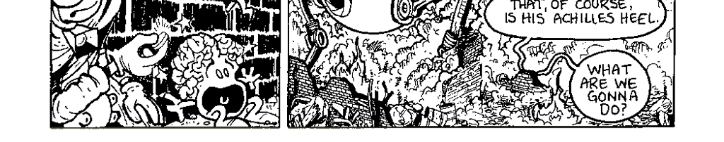


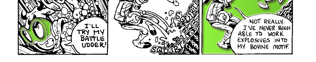


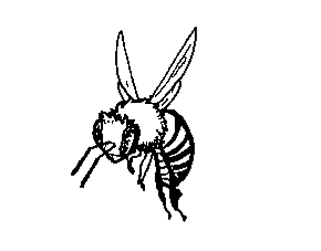
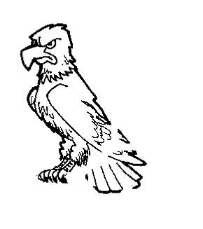
The Birds and the BeesLet me tell you about the birds and the bees - or their eyes, at least. Like many animals, birds and bees depend on vision to navigate, avoid being eaten, find food and choose mates, but, they accomplish these things with two very different types of eyes. You don’t have to be a trained naturalist to differentiate between the compound eye of the common honey bee Apis mellifera and the massive yellow camera eyes of a bald eagle. Despite their differences, both eyes paint effective pictures of the world for their owners: |
 |
|||||
Eyes Run in Our FamilyBiologists classify organisms based on how they look and their genetic similarities. Eagles are birds and birds belong to a group known as the vertebrates. Vertebrates have internal skeletons and a series of bones called vertebrae that run down their back and encase their spinal cord. You are a vertebrate. Assuming the vertebrate in question is not a blind cave fish or cave salamander, vertebrates have camera eyes, although the design of each varies based on the species’ evolutionary history and ecological circumstances. Honey bees are invertebrates. Invertebrates are multicellular animals without a backbone. This is a tremendously large group of animals that ranges from microscopic worms to the whalesized giant squid! Insects belong to a sub-category of invertebrates known as the arthropods. The arthropods are classified as having external skeletons, jointed legs and no backbone. Arthropods have compound eyes which can vary in size, shape and complexity depending on the ecological circumstances and lifestyles of the arthropod in question. When we consider the position of birds and bees on the family tree of life, we see that the branch that led to vertebrates split more than 500 million years ago from the branch that eventually led to arthropods. The common ancestor of these two lines probably did not have much in the way of visual apparatus. Thus, members of each line evolved their elaborate visual devices independently. Compound eyes and camera eyes represent different solutions to the same ecological challenge: how to perceive light. Consequently, although they may not look very much alike, they share many of the same functional characteristics. |
 |
|||||
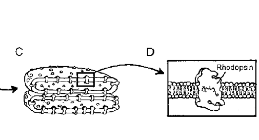
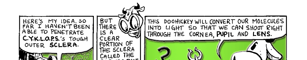
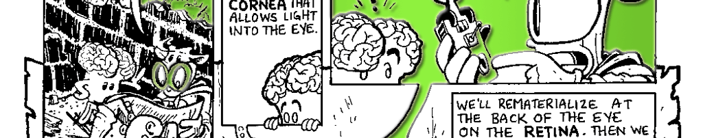
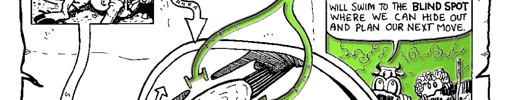
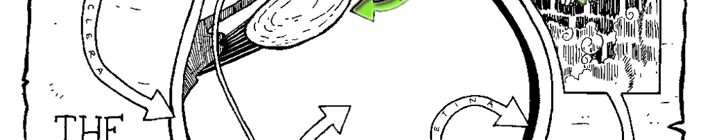
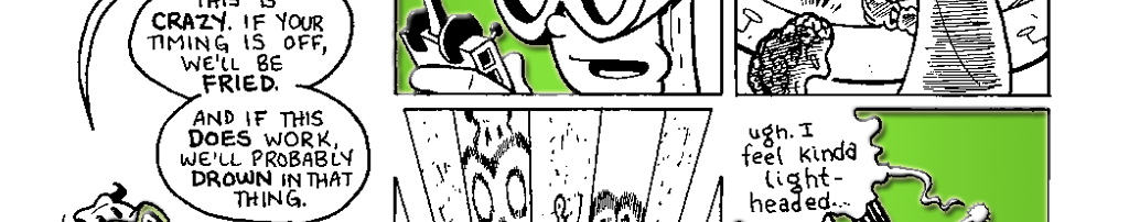
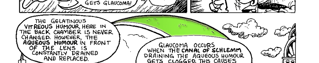
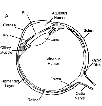
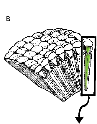
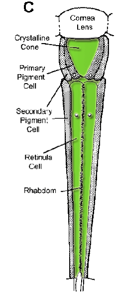
Let the Sunshine InAn eye’s first job is to let the sunshine in. Most places on an animal’s body are hard to see through, so for light to penetrate (and eventually be absorbed) there needs to be a transparent window someplace. In vertebrates, this region of the eye is called the cornea (Fig. 3.1 A) and in insects it is called the corneal lens (Fig. 3.1C). The “white of your eye” is a fibrous and relatively rigid structure called the sclera that gives the eye its shape. The cornea is a region of the sclera that has become transparent, in large part because it contains no blood vessels. Because of this, corneal transplants are the most successful type of transplant because the immune system has no way to reach the new cornea and reject it. While the eye of an eagle is a single optical unit with only one cornea, the compound eye of a bee is composed of thousands of optical units called ommatidia, each with its own comeal lens (Fig. 3.1B-C). The corneal lens is a modified region of the bee’s rigid exoskeleton that is devoid of most of its pigmentation. Iris is a Very Good PupilIn both birds and bees, once light has passed through the cornea or comeal lens, it must pass through another clear structure that acts, to a greater or lesser extent, to focus the light. Light in the insect compound eye passes from the comeal lens immediately to the adjoining crystalline cone (Fig. 3.1C). In conjunction with the corneal lens, the crystalline cone bends incoming light and focuses it on the rhabdom at far end of the ommatidium. The camera eye of a vertebrate also has a second focusing structure that works in conjunction with the cornea. However, they are not in direct physical contact. After light enters the eye through the cornea, it travels through the aqueous humor fluid of the front chamber of the eye and passes through the hole in the middle of the eye called the pupil. The pupil is formed by a circular muscle called the iris and can open and close to control the amount of light entering the eye. In the dark, the pupil widens to allow as much light in as possible and in bright light it does the opposite. To see this happen, try the following experiment. Sit in the dark with a friend for a minute of two. Once your eyes have acclimated, shine a flashlight in your friend’s eyes and watch his or her pupils contract. (You should probably warn your friend that you are going to do this or they might not be your friend for long.) Once through the pupil, the light hits the oval-shaped lens. Traditionally we think of the lens as the structure that bends incoming light and focuses it on the retina at the back of the eye. However, the effectiveness of the lens in bending light depends on where you live. |
||||||
  |
||||||
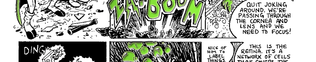
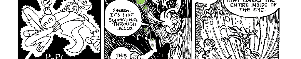
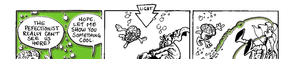
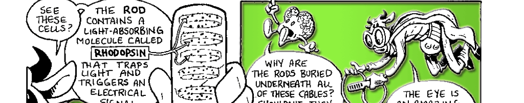
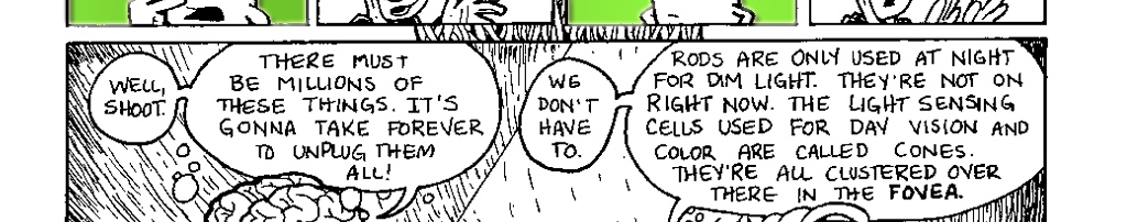
I'm Having Trouble FocusingIn aquatic vertebrates like fishes, the lens is the primary means by which light is focused, but in terrestrial organisms like us, the cornea does most of the work. To understand why this is the case, it is important to bear in mind three important things: 1) the space between the cornea and lens is filled with a fluid (aqueous humor), 2) fluids are denser than air and 3) light travels faster through air than it does through water or a transparent solid like a lens. When we see something, light has traveled through the air to reach our eyes at a speed of 186,282.397 miles per second. When a wavefront of light passes through the curved surface of the cornea, two things happen. First, it slows down to 140,000 miles per second as it moves from the air into the much denser aqueous humor. Second, the light is bent and focused by the curved surface of the denser medium. This bending of light is called refraction. Things are different for fishes. Light reaching their eyes passes from one fluid (the water) to another (the aqueous humour). This fluid-to-fluid transition doesn’t slow the light and thus, doesn’t bend or focus it much either. Consequently, fish rely upon the transition from the aqueous humour to the solid lens to slow the light and do most of the bending. Fish lenses slow light down to 124,000 miles per second. Because the lens is so thick, the light spends a relatively long time slogging through it and gets significantly refracted. By comparison, terrestrial vertebrates tend to have smaller, flatter lenses that are just responsible for fine-tuning focus. You're Not Going AnywhereOnce the light is bent, it is focused on a structure where it can be absorbed and trigger a signal to be sent to the brain. In bees, the light in each ommatidium is focused on an area called the rhabdom, a place where the fingers of several neighboring retinula cells overlap (Fig. 3.1C). In an eagle, the light is focused on a layer of cells lining the back of the eye called the retina (Fig. 3.1 A). The cells of the rhabdom and retina both contain special molecules called photopigments that can absorb a photon of light. Once absorbed, the photon’s energy causes the photopigment to change shape and activate a series of chemical reactions that will lead to an electrical change in the cell. Of course, photons are quick little buggers and not every incoming photon hits a photopigmcnt. Both the compound eyes and camera eyes have additional pigments in place to absorb or redirect these stray photons, preventing them from bouncing around inside the eye and blurring vision. In the compound eye, each ommatidium is lined by pigment cells that channel photons toward the rhabdom and prevent light leakage between the ommatidia. The vertebrate eye has a layer of heavily pigmented cells behind the retina that lap up what is missed by the photoreceptive rod and cone cells. |
|
|||||
|
||||||
 |
|
|||||
Try To See It My WayCamera and compound eyes aren't the only way to see the world, of course. Consider the figure of eye evolution from Nilsson and Pelger (Fig. 2.2). At one end of that continuum, we see the flat patch of photosensitive cells found in animals like flatworms, segmented worms and some crustaceans. At the other end of the spectrum, we have the camera eyes found in vertebrates, cephalopods (like squid and octopuses) and some jellyfish. And in between are a number of visual intermediates capable of forming images of varying complexity. These intermediate eyes are found throughout the animal kingdom and in a variety of ecological niches. The functions of the visual intermediates vary. Flat and cupulate eyes can be found in worms, arthropods and vertebrates. They don’t form images but they can be used by organisms to orient toward or away from light (a useful skill, if you are trying to avoid the bright open spaces filled with big, hungry predators). They may also be used to gauge day length, which can be useful in coordinating feeding and breeding behaviors. Pinhole eyes are an elaboration of the cupulate eye. They contain a small aperture that can be used to focus light and work on the same principle as a pinhole camera. The small hole in the eye bends and focuses light to form an image on the photosensitive cells in the eye. The smaller the hole, the better the image resolution. But there is a trade-off to this approach: as the hole gets smaller, the image gets darker. Eventually, selection for additional increases in resolution (by making the hole smaller) is no longer favored because of lost image brightness. These eye can be found in ribbon worms, some arthropods and the ancient Nautilus. There are several solutions to the ecological challenge of perceiving light. A survey of eyes in nature clearly demonstrates that it is incorrect to assume that the only eye worth having is an eye like ours. Natural selection does not work to make organisms more like us. It acts to preserve adaptations organisms need to survive in their environment. If a sedentary clam doesn’t need an elaborate eye to sit and filter feed, then the economics of evolution will favor something simpler. Likewise, a predaceous squid will need far more sophisticated eyes to hunt at high speeds. And there are costs to having an eye. Eyes may not really be the gateway to the soul, but they can be a gateway for diseases. They are also vulnerable to predators. Under an organisms’ unique environmental conditions, the costs of building an eye must be weighed against the needs of the animal. For your consideration
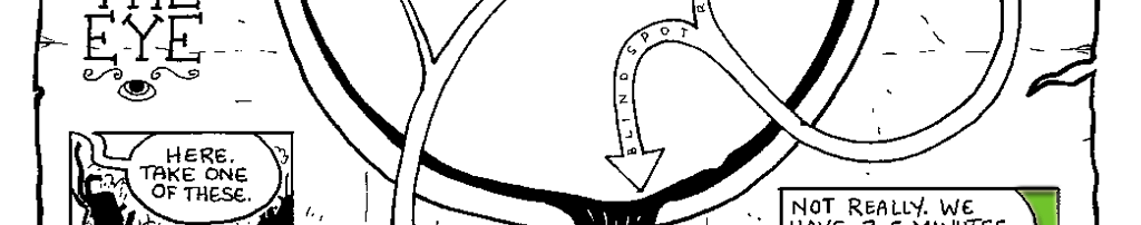
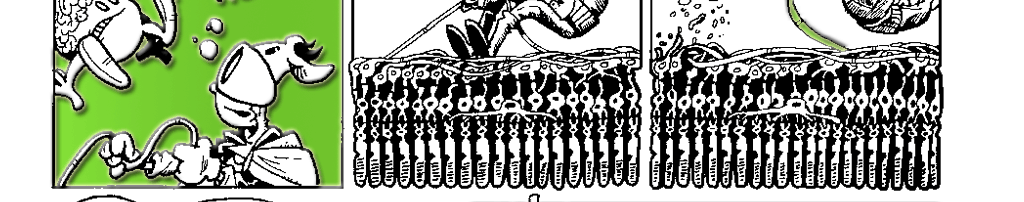
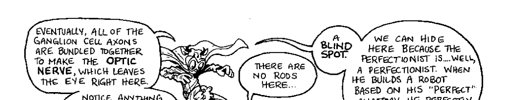
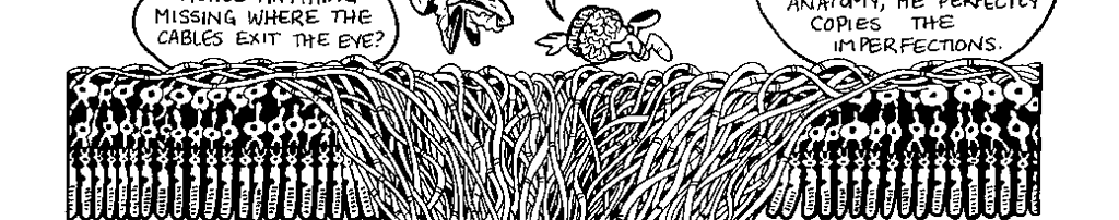
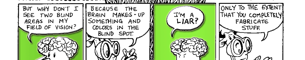
Blind Spot ActivityClose your left eye and stare at Wrinkles with your right eye. Now move the page closer to your face keeping your right eye on Wrinkles the whole time. When the page is about a foot from your face, the magic eye should disappear. It will reappear if you continue moving the page closer to your face. Wha' Happened?As you move the page closer to your face, you eventually positioned the magic eye on the blind spot of your right eye. Since there are no rods and cones there, the image doesn’t stimulate your retina. Your ever resourceful brain looks at the stimulation all around the blind spot and fills in the gap. Since the magic eye is surrounded by white, the brain colors in the area with white and the magic eye appears to disappear. |
||||||